
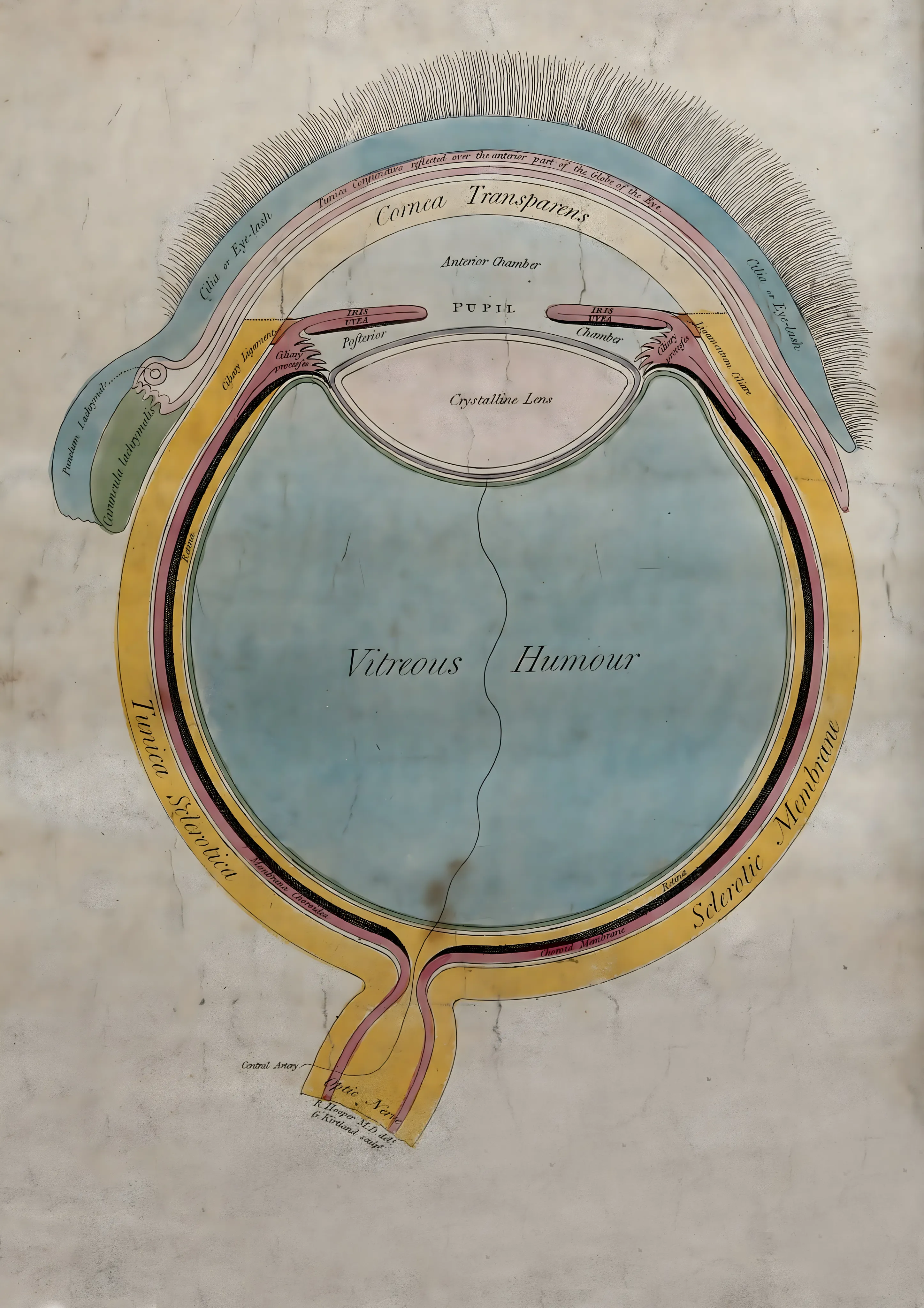
Anatomical Diagram of the Human Eye
Anatomie
FromNow €5.00Was €6.00
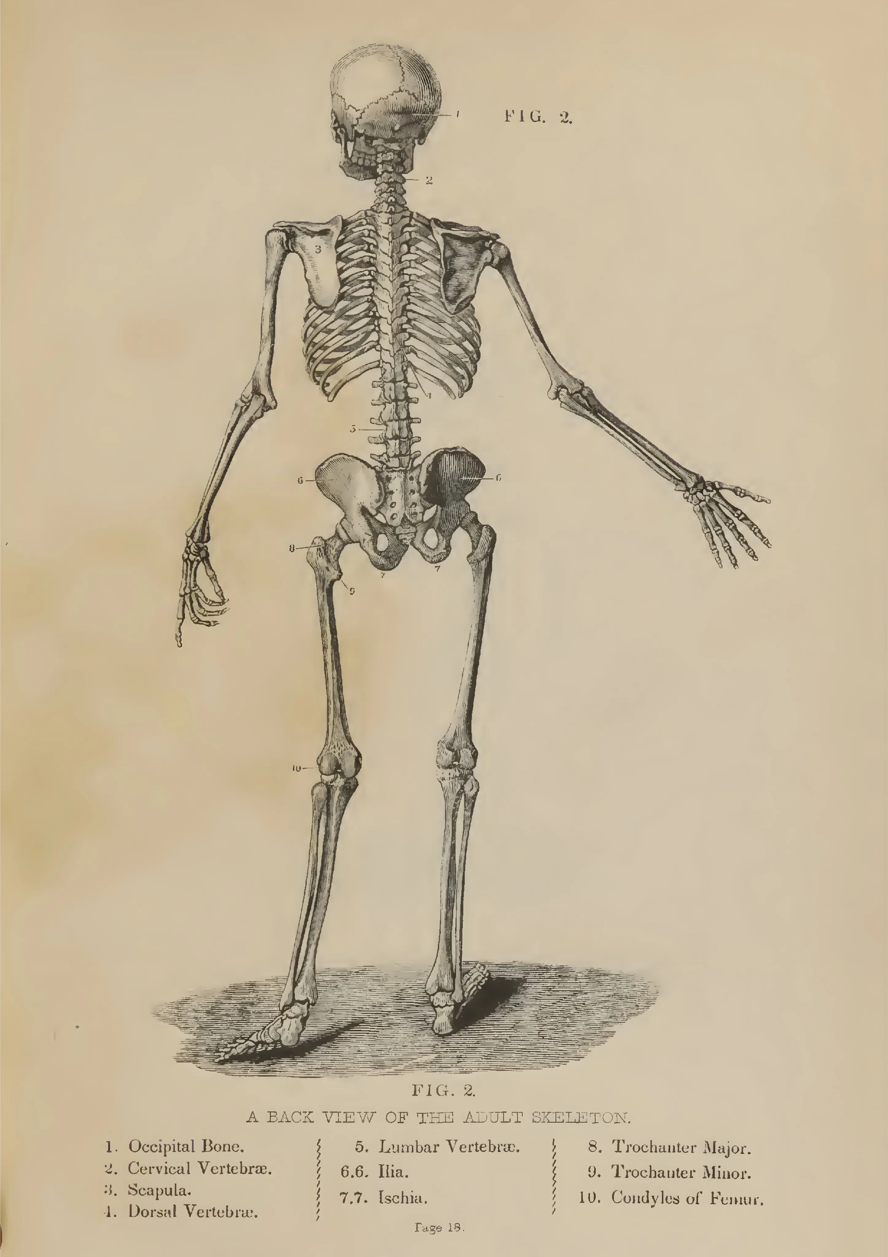
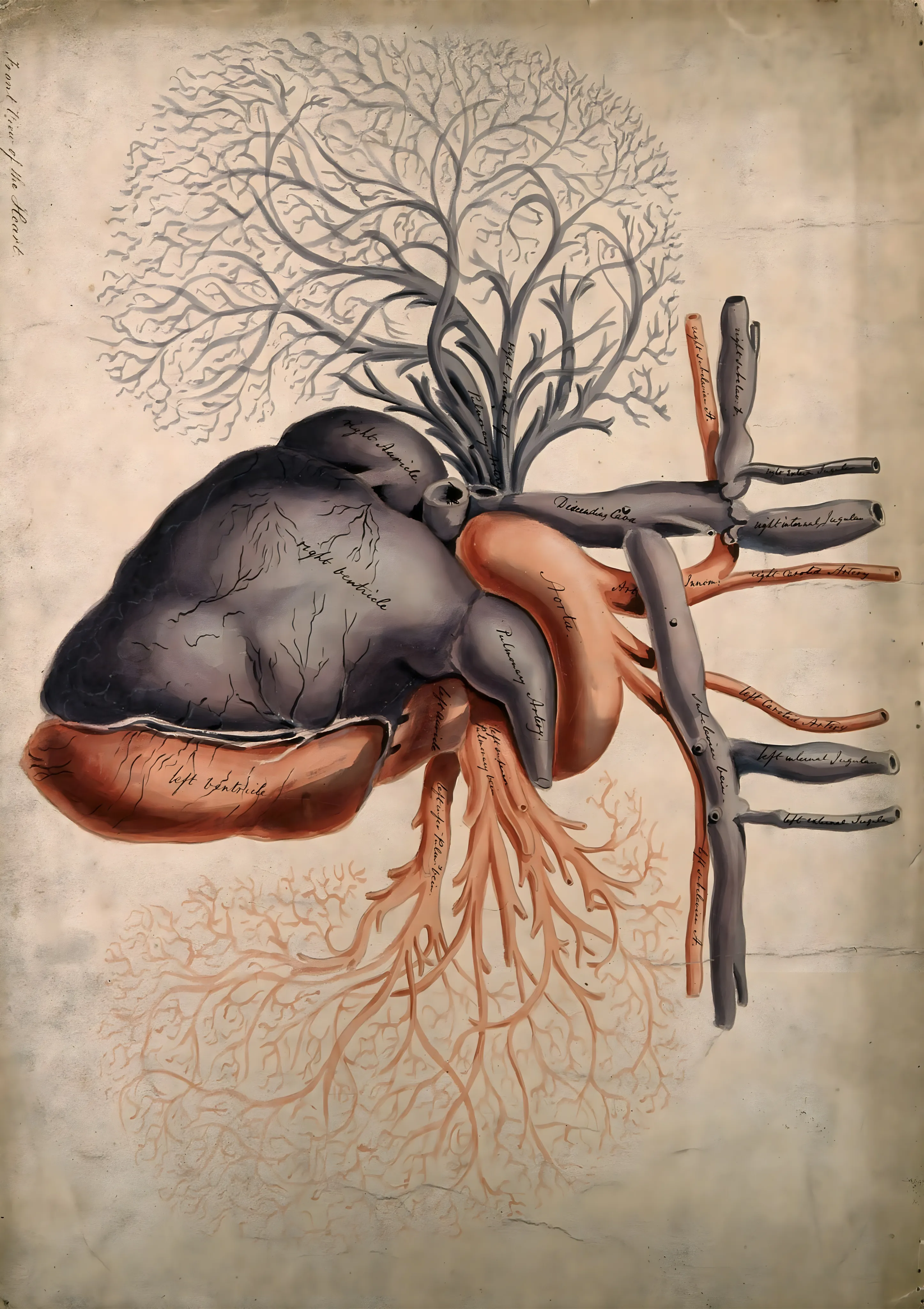
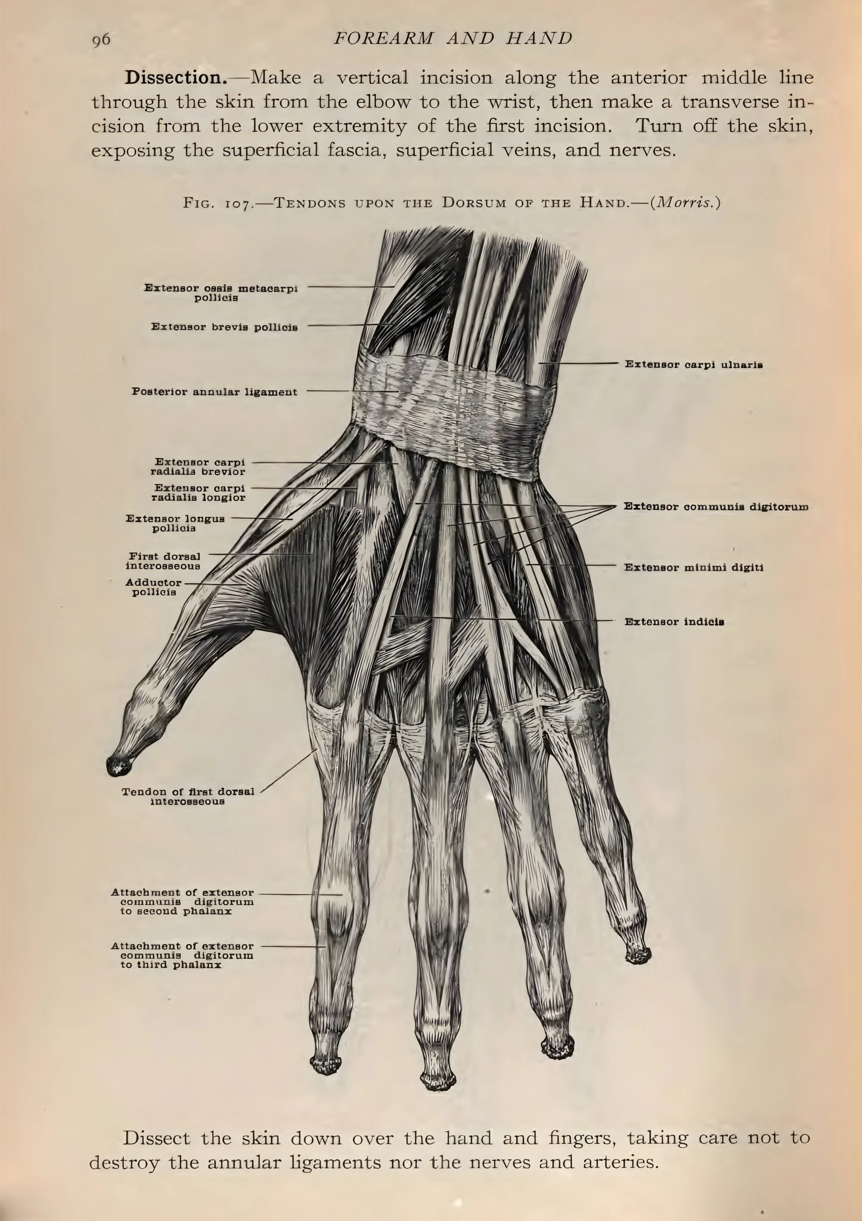
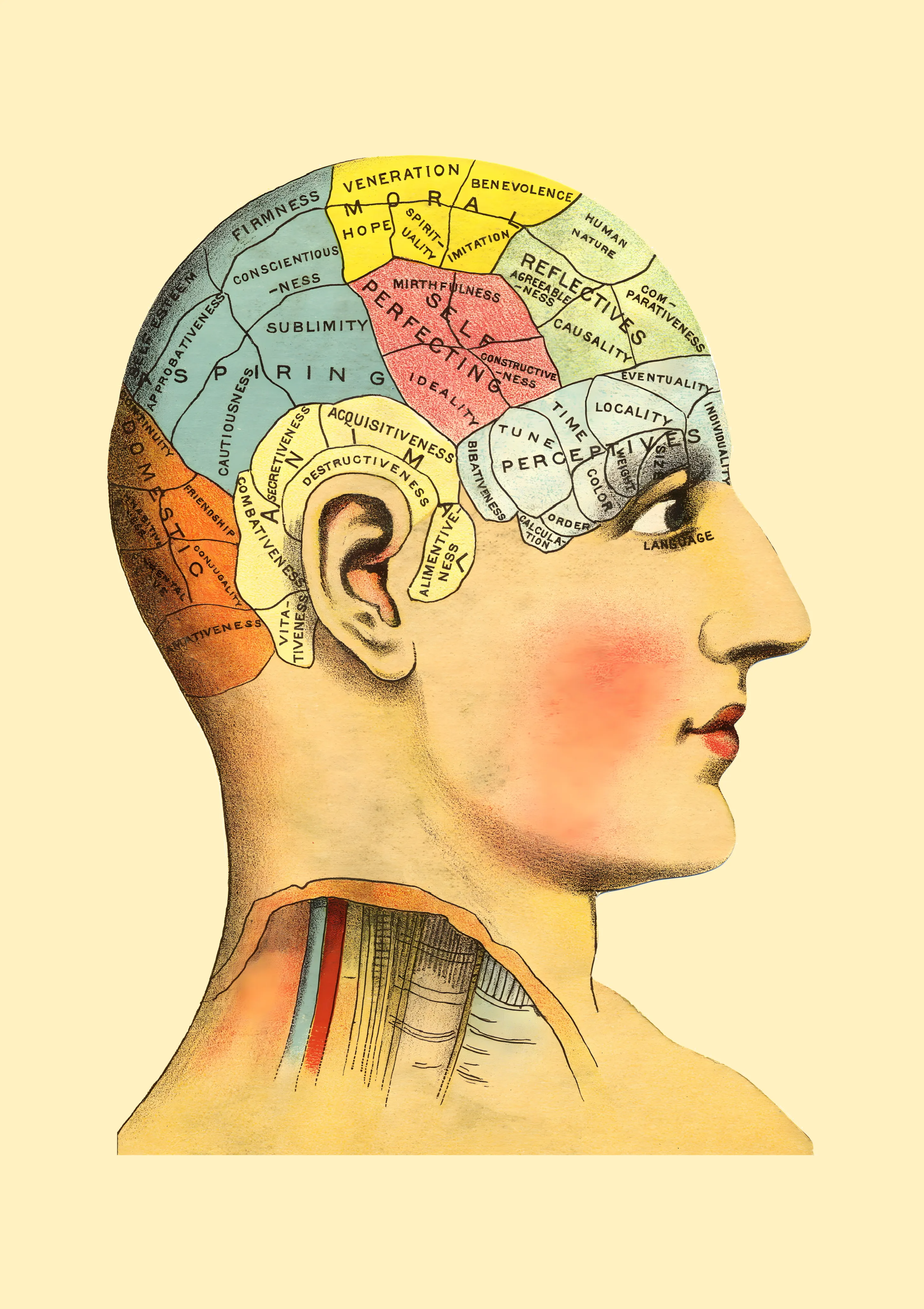
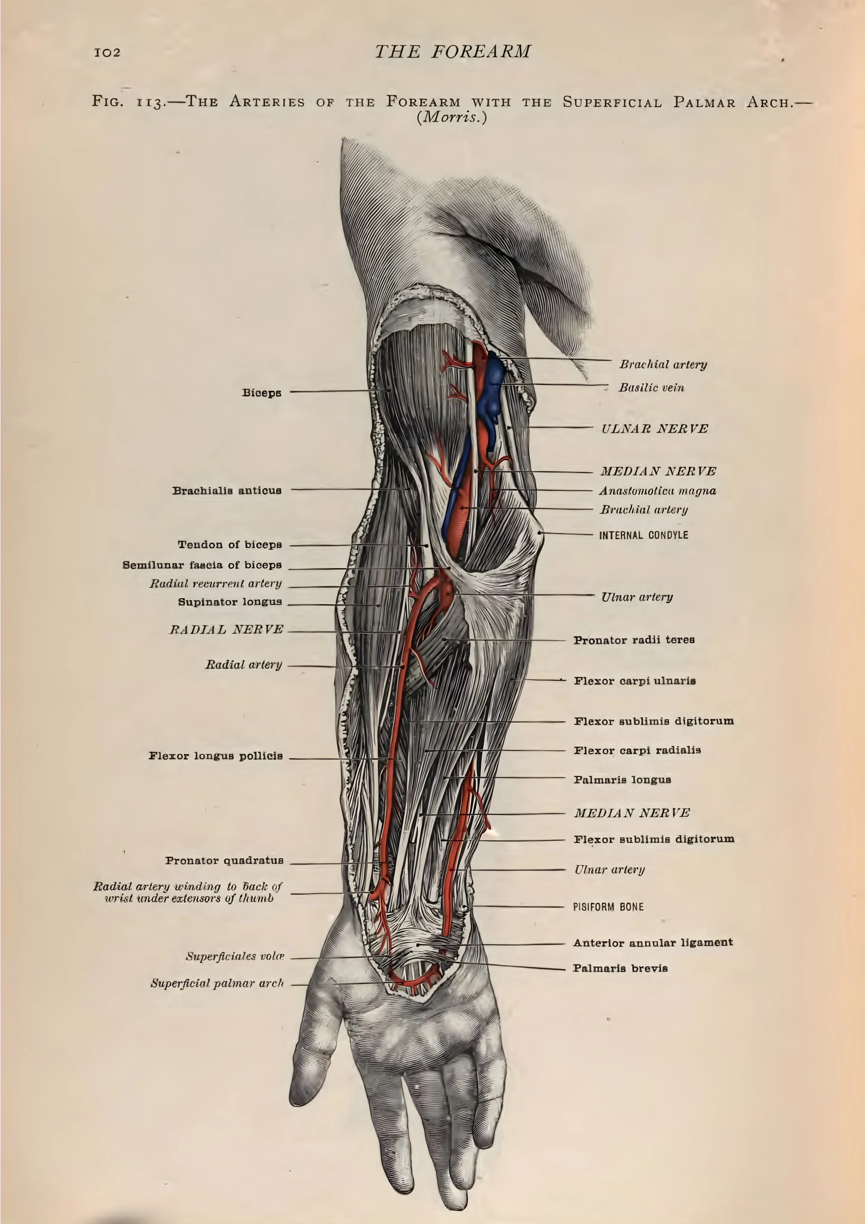
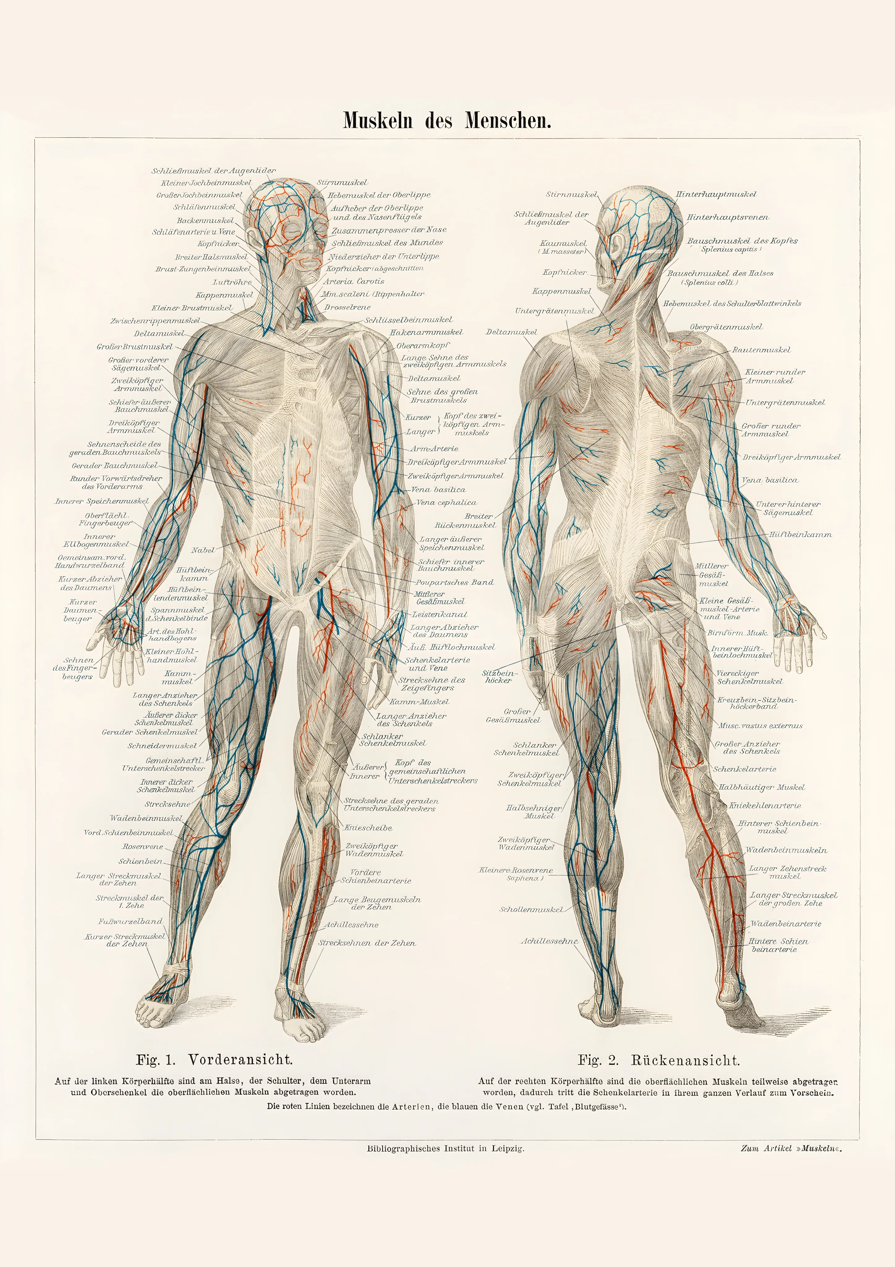
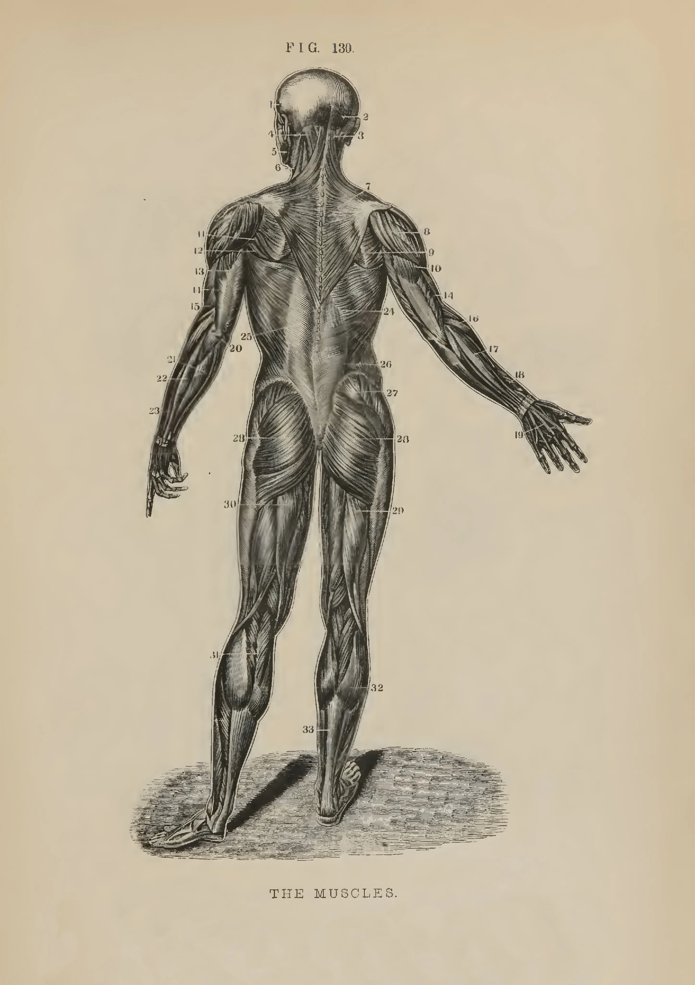
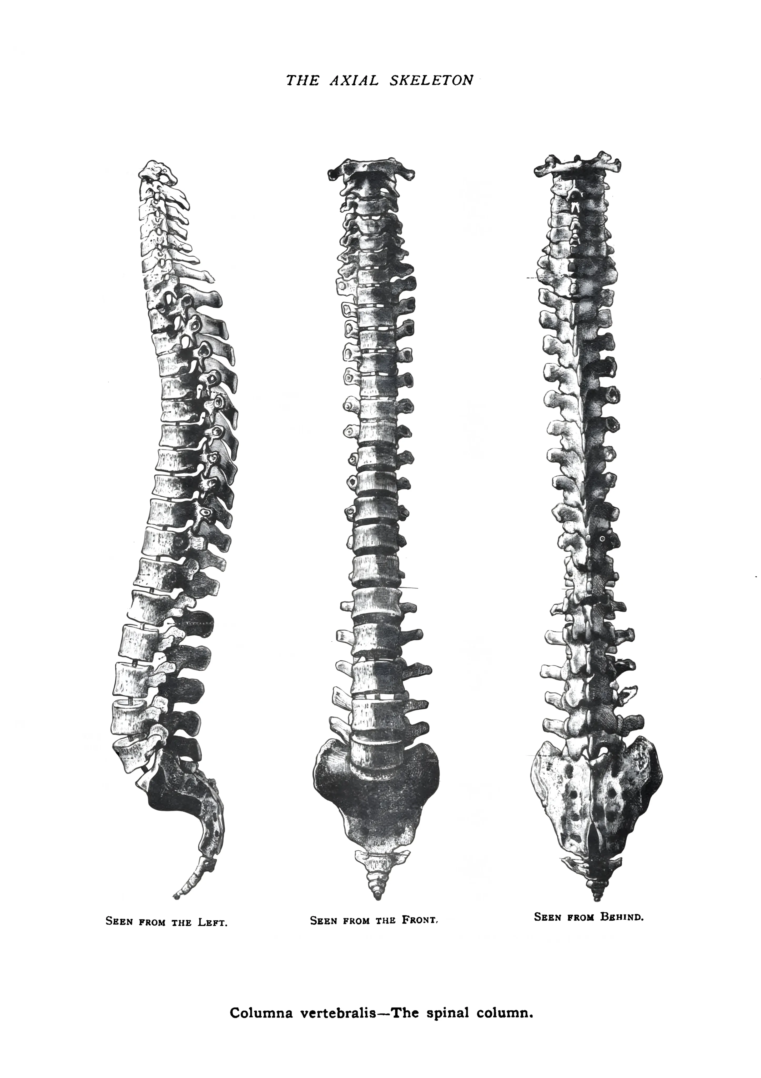
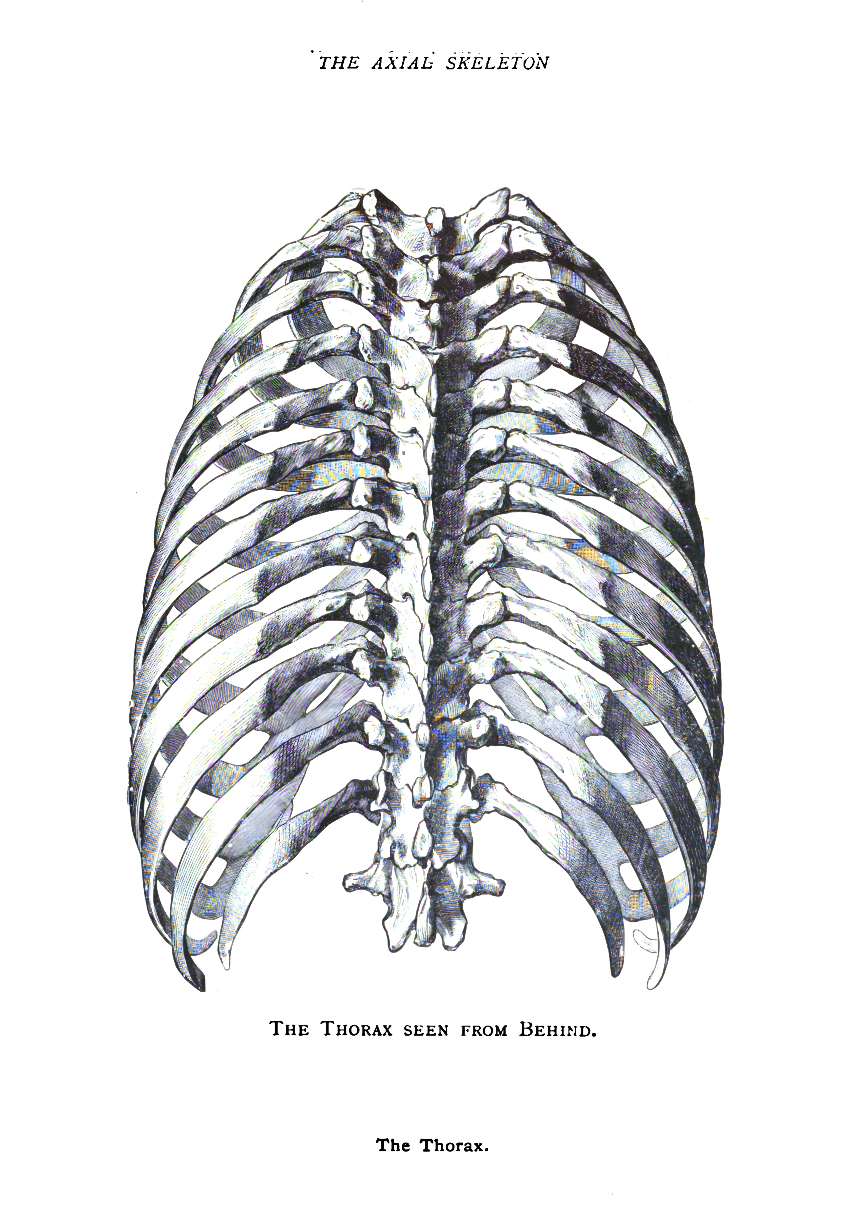
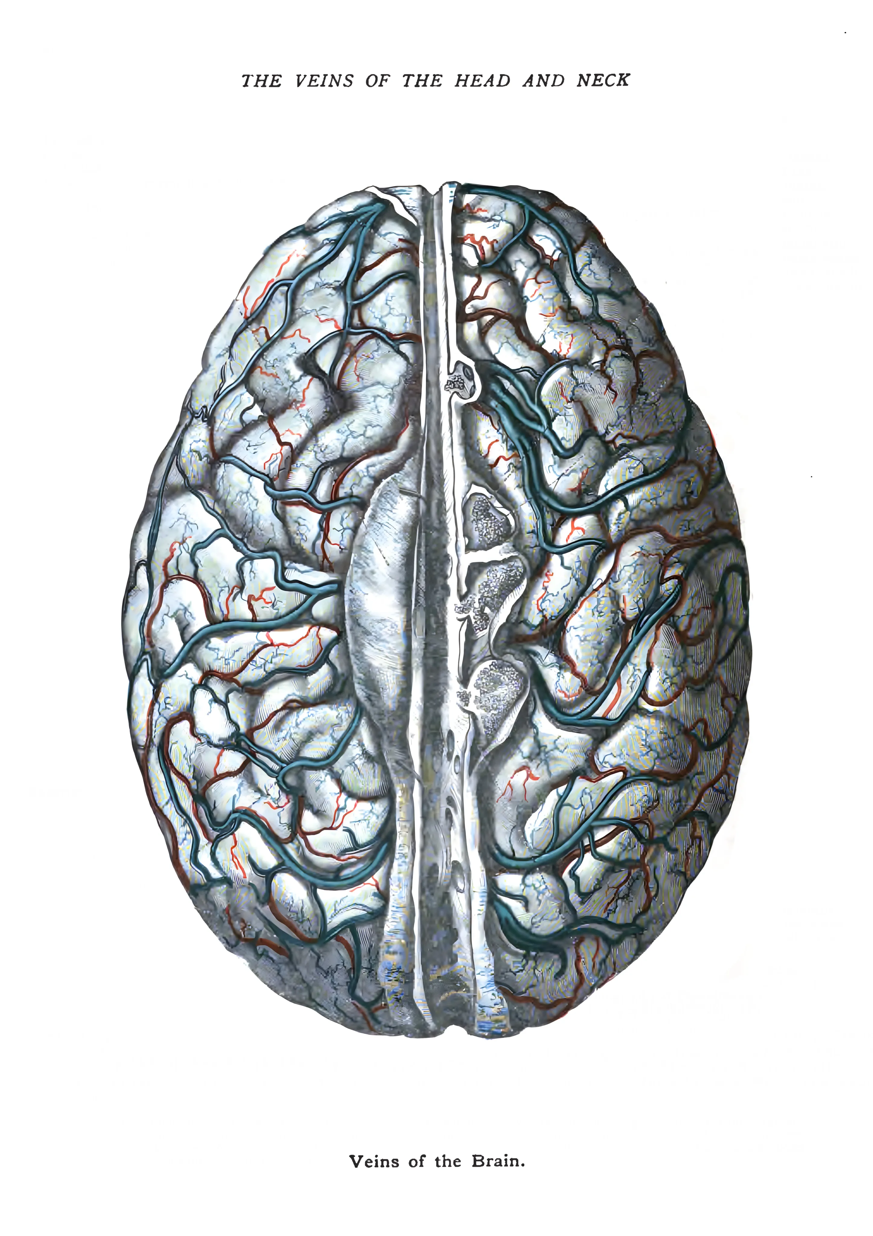
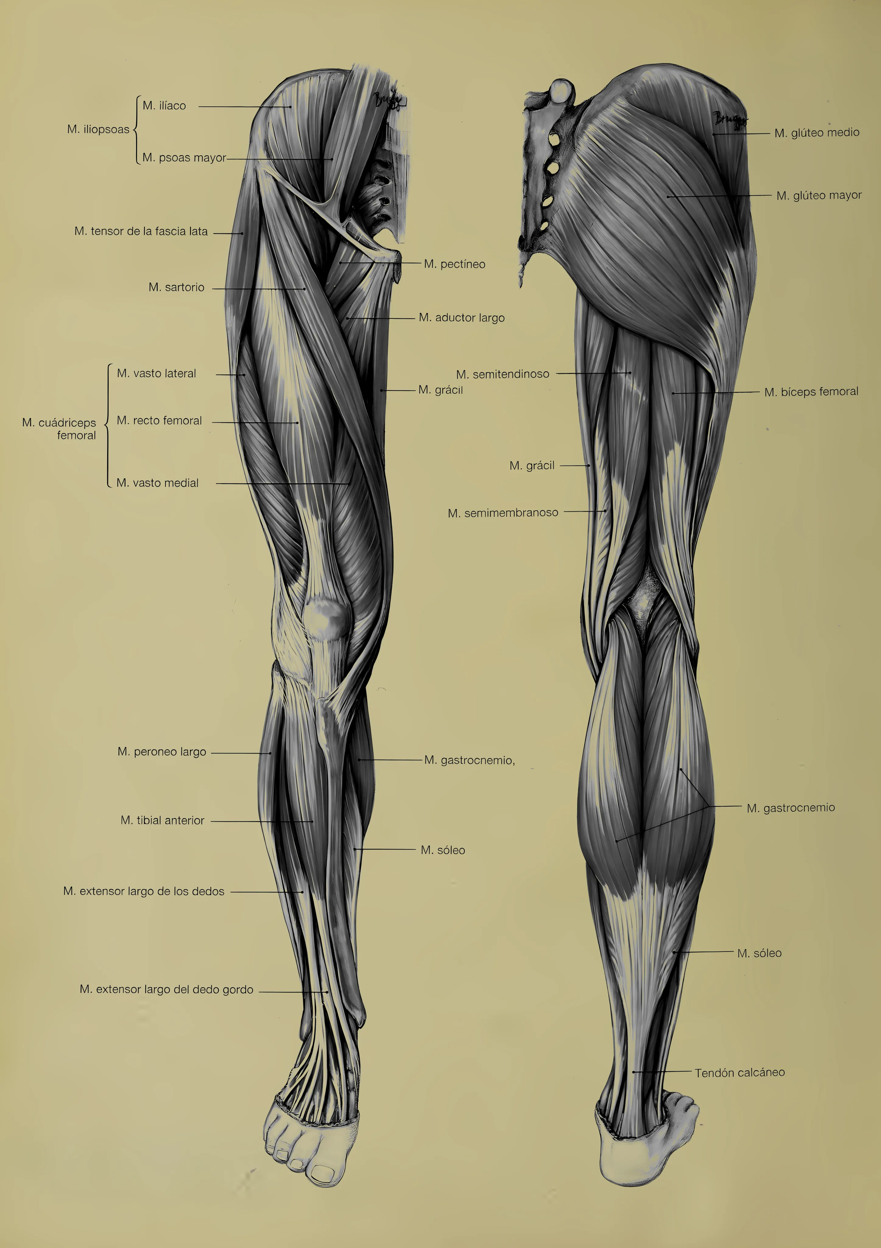
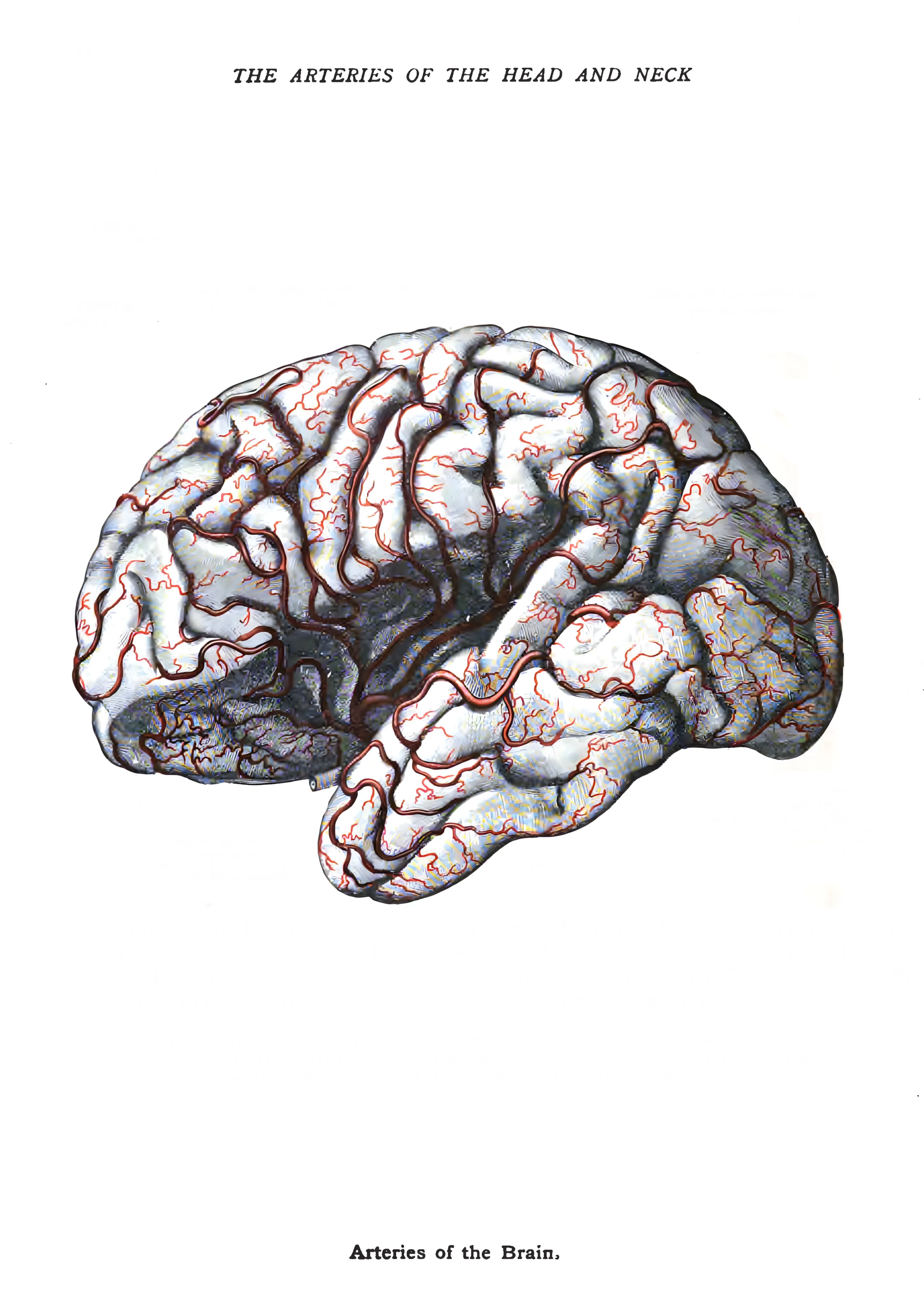
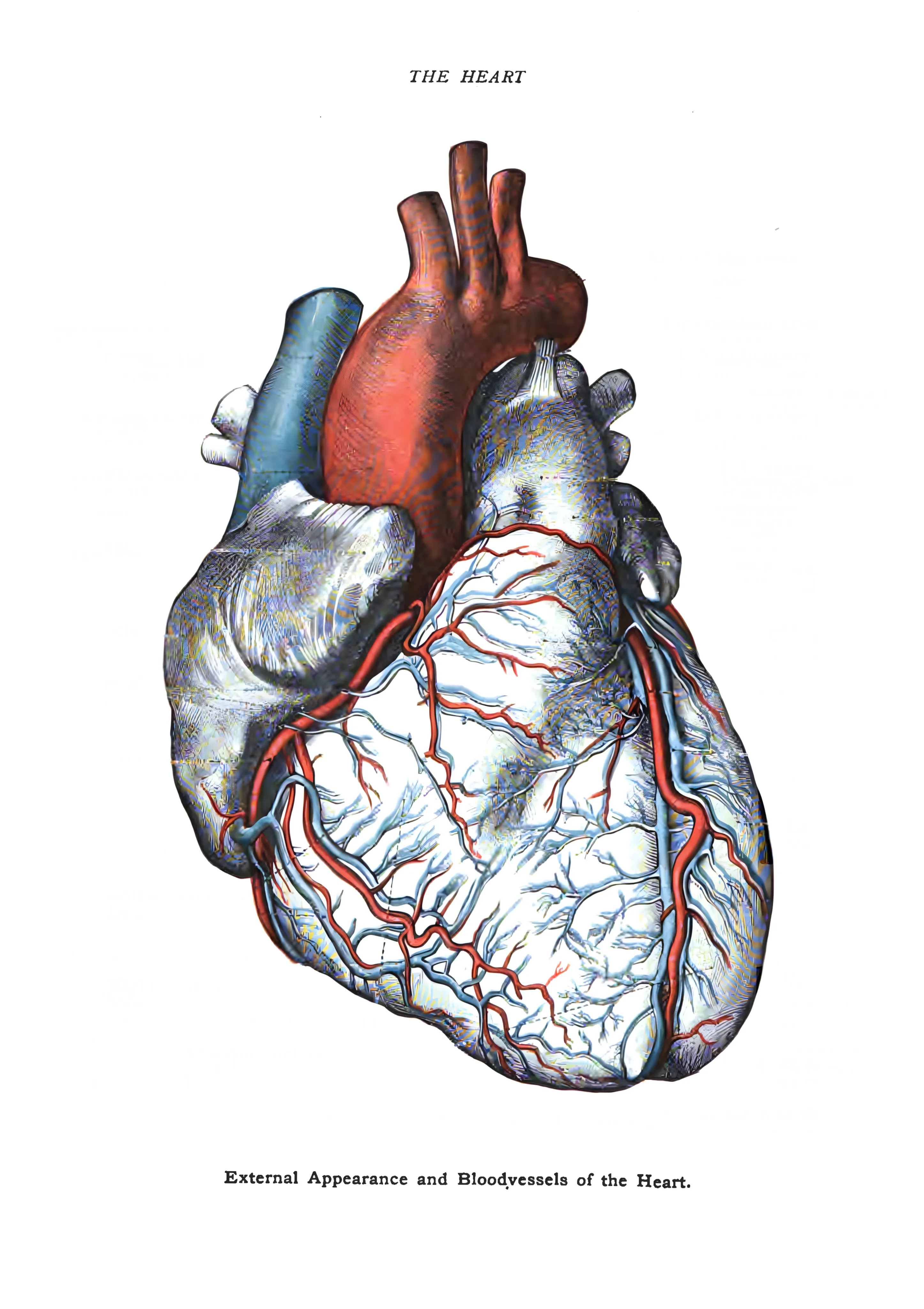
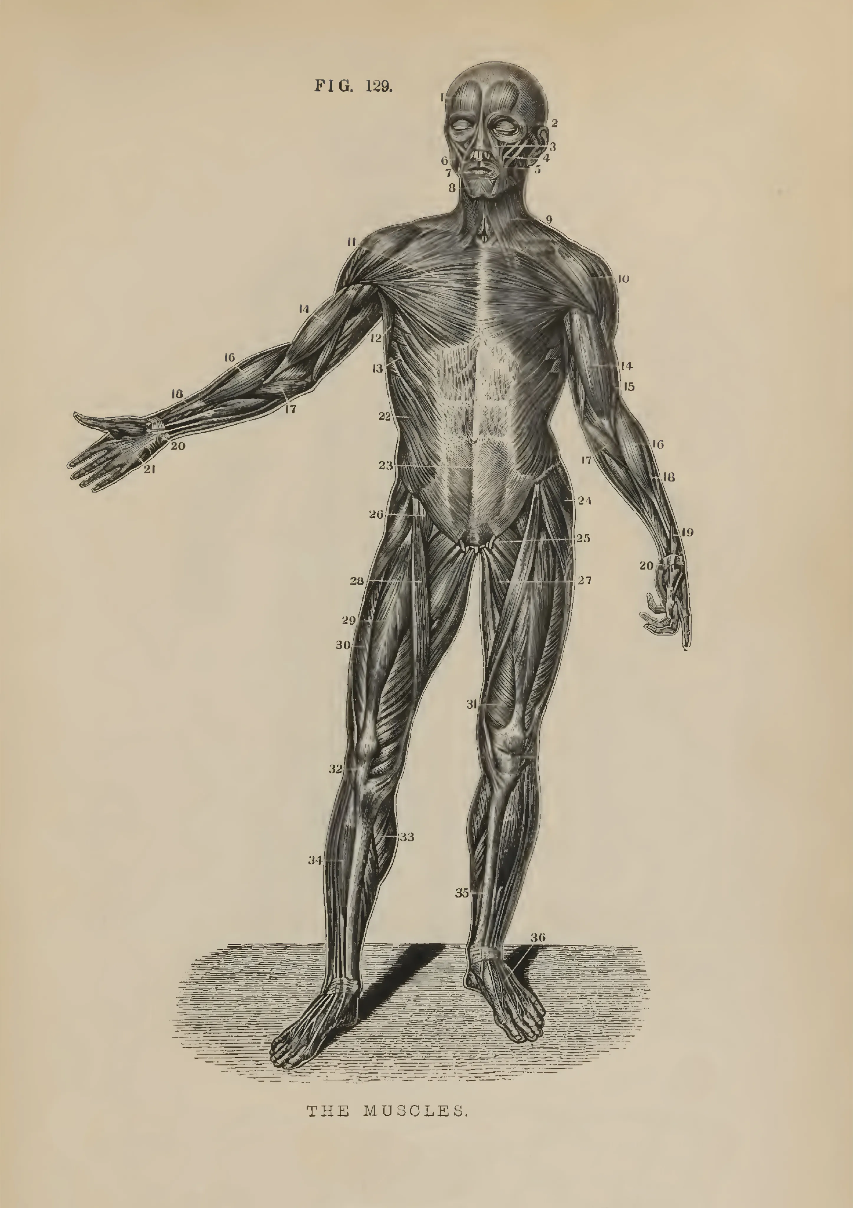
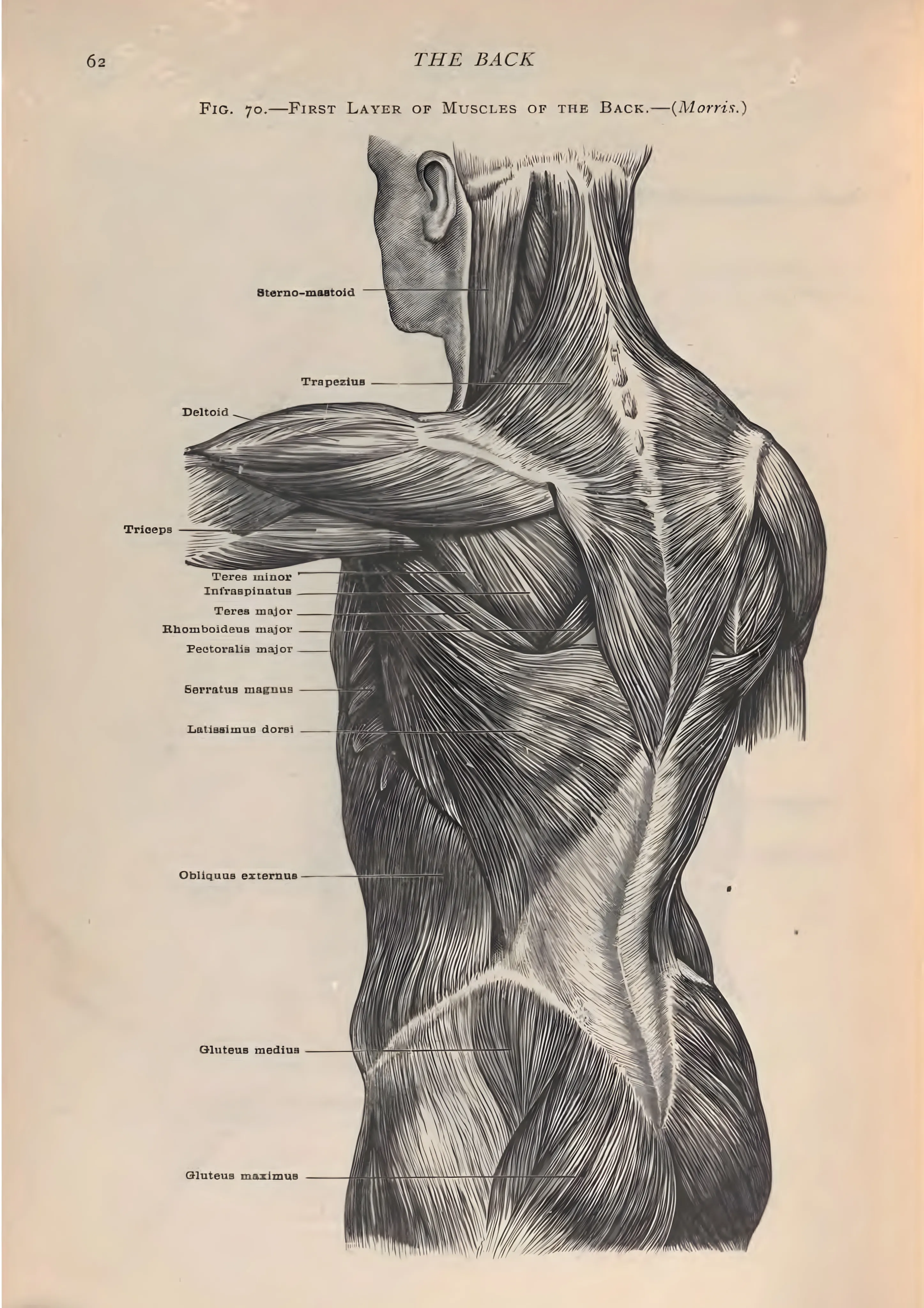
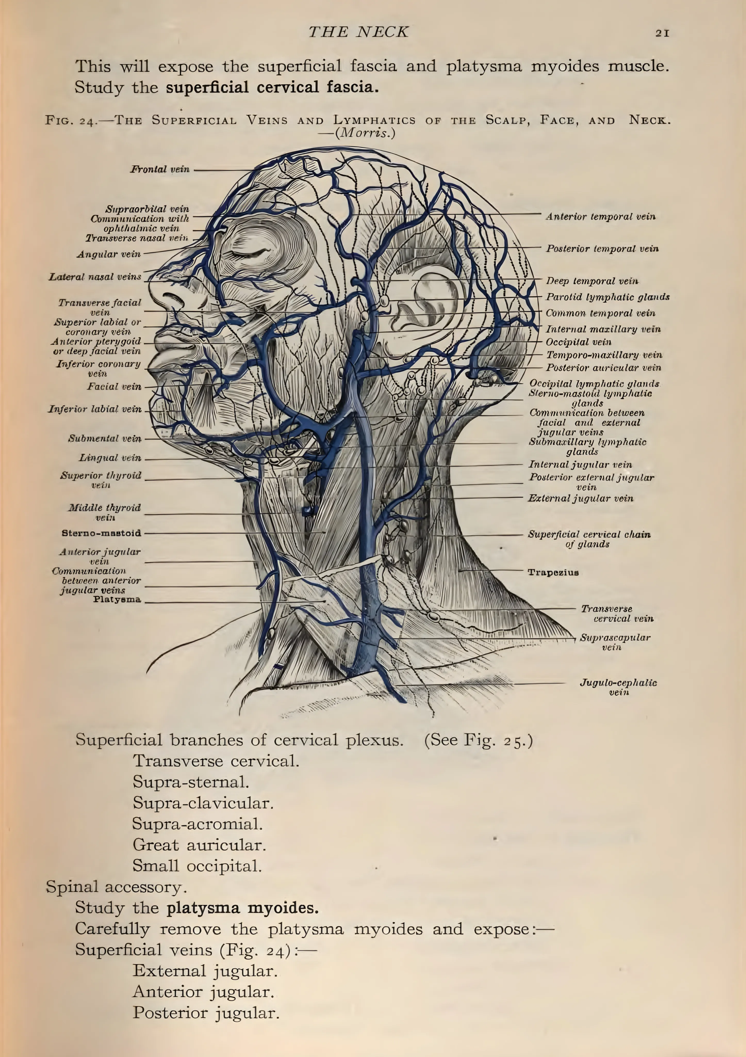
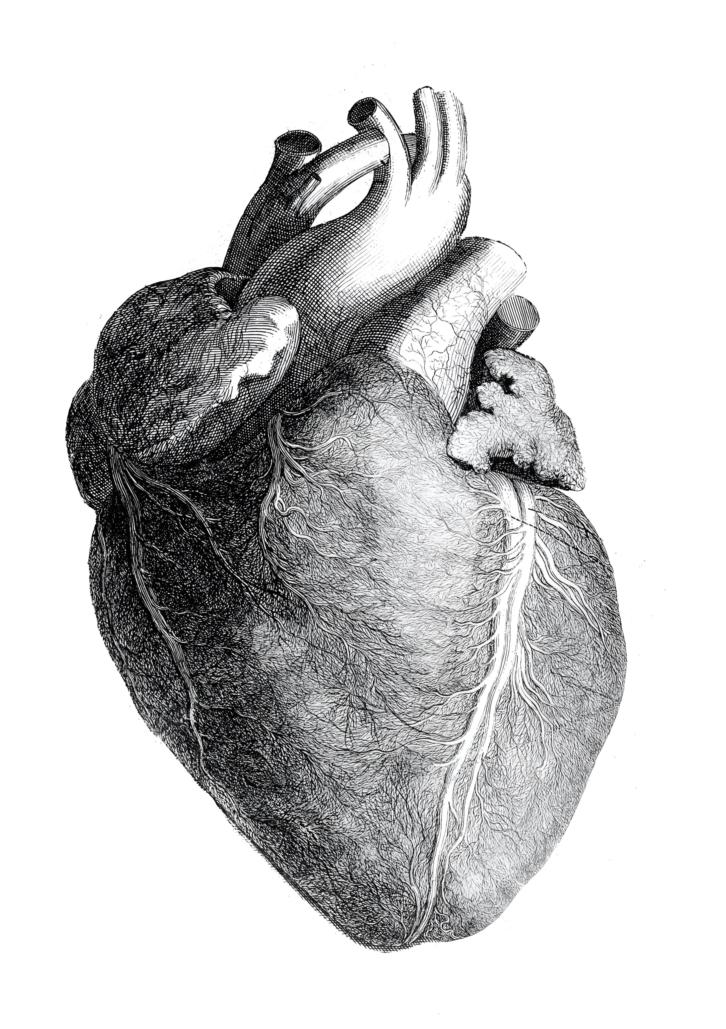
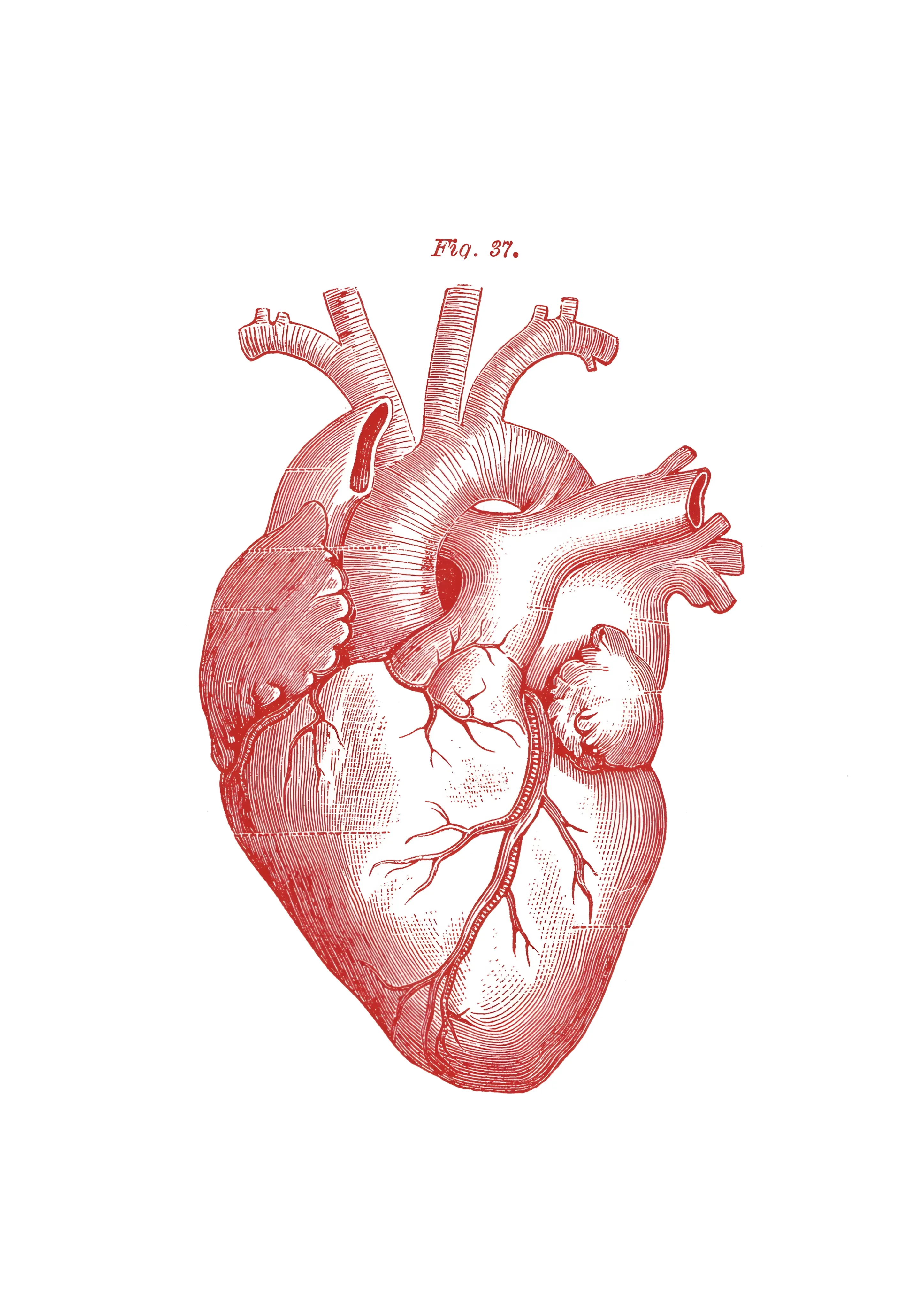
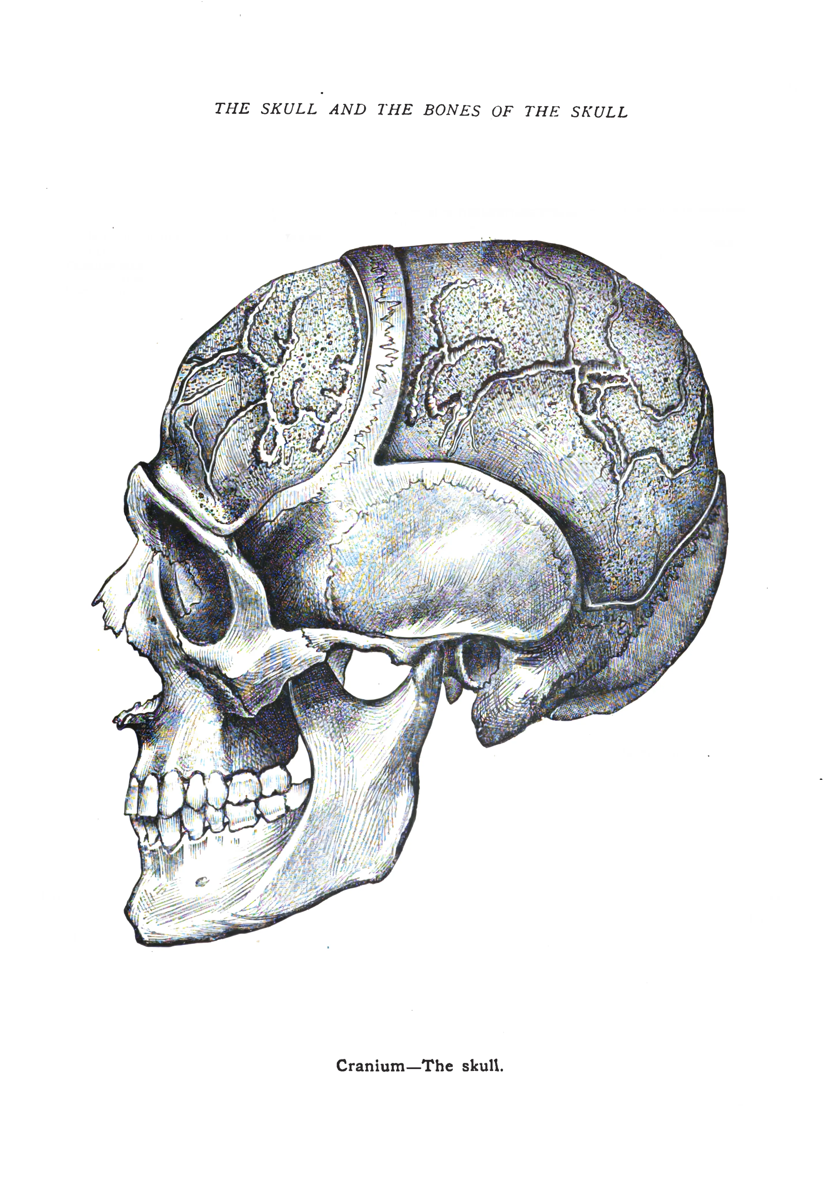
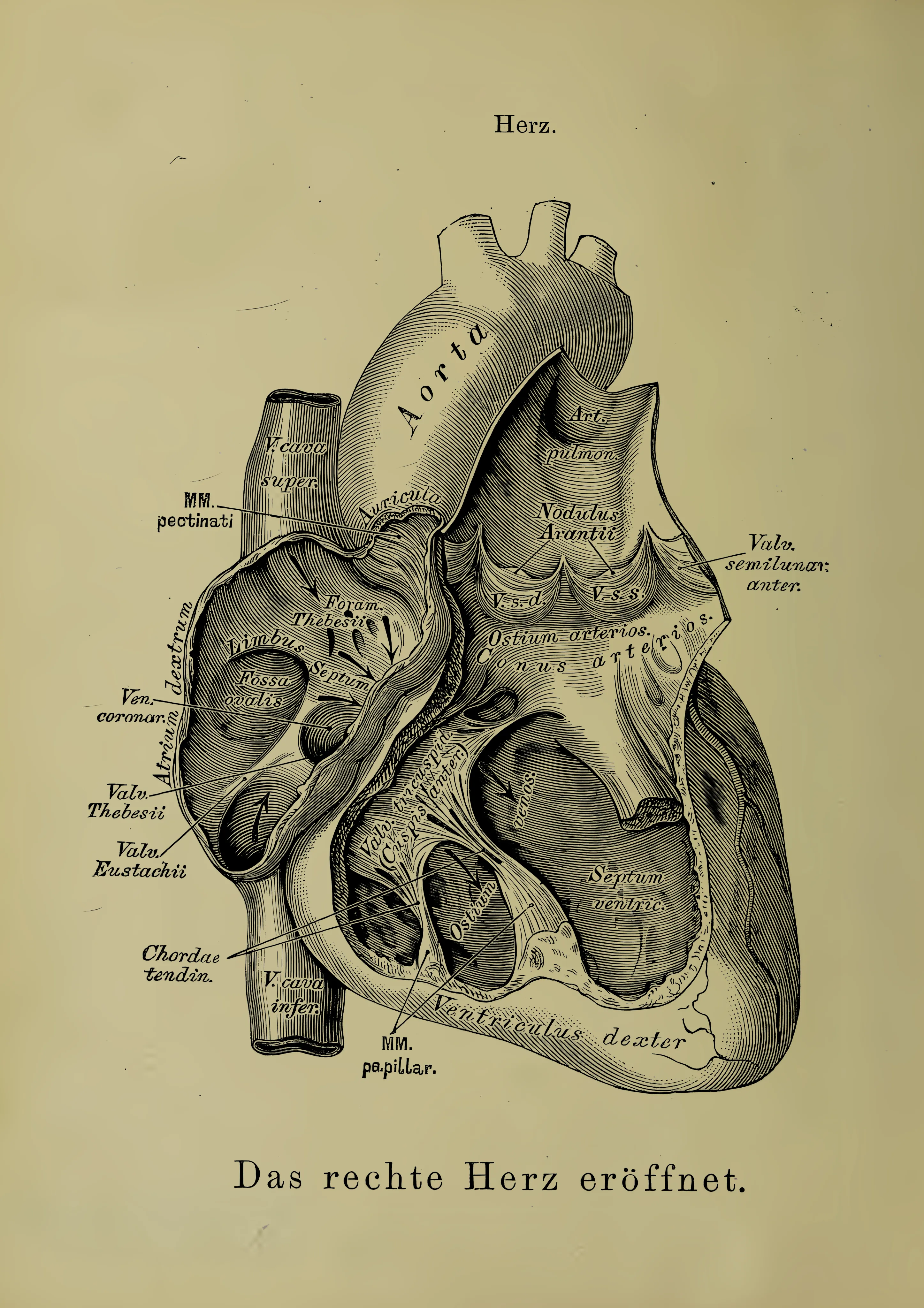
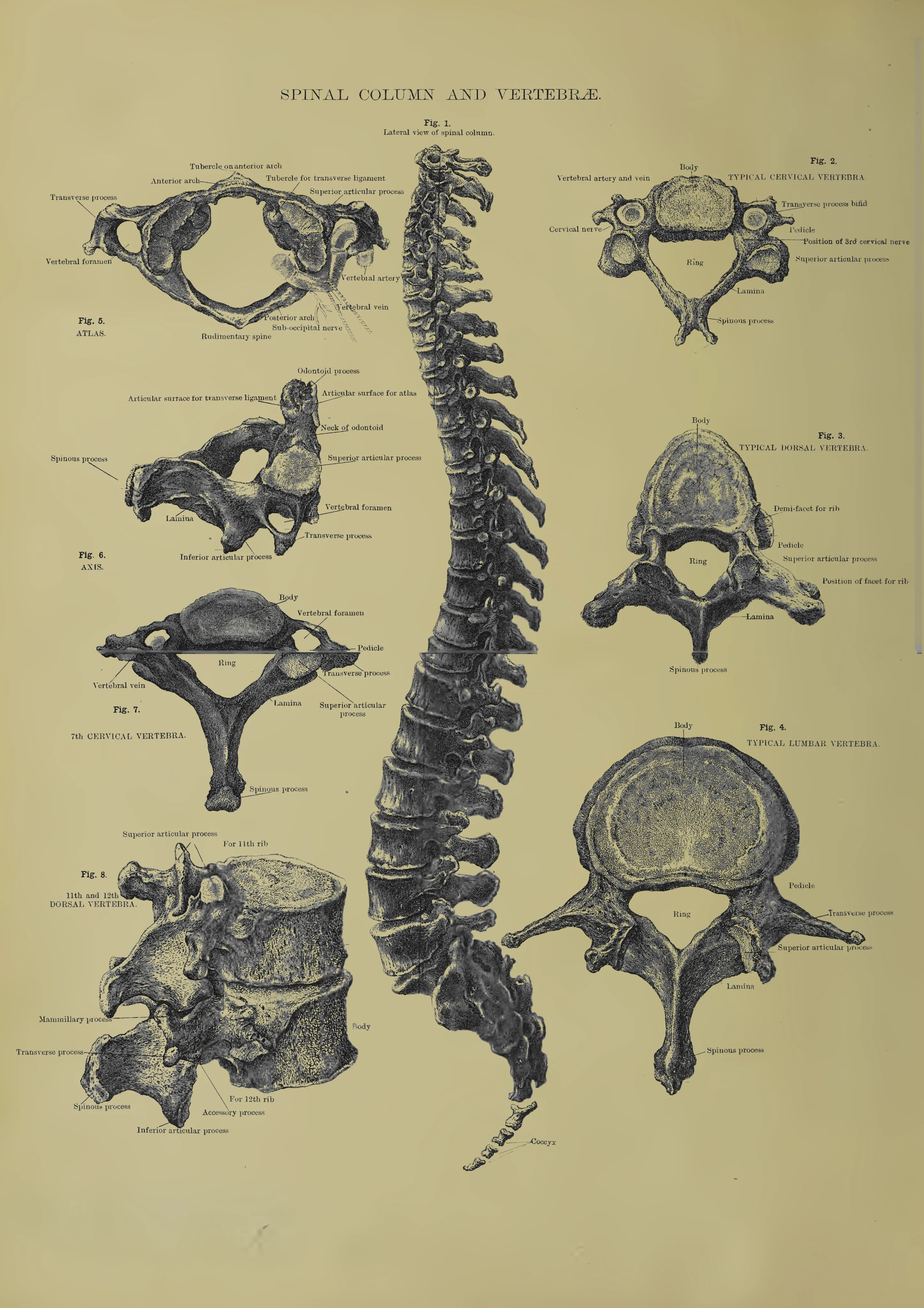
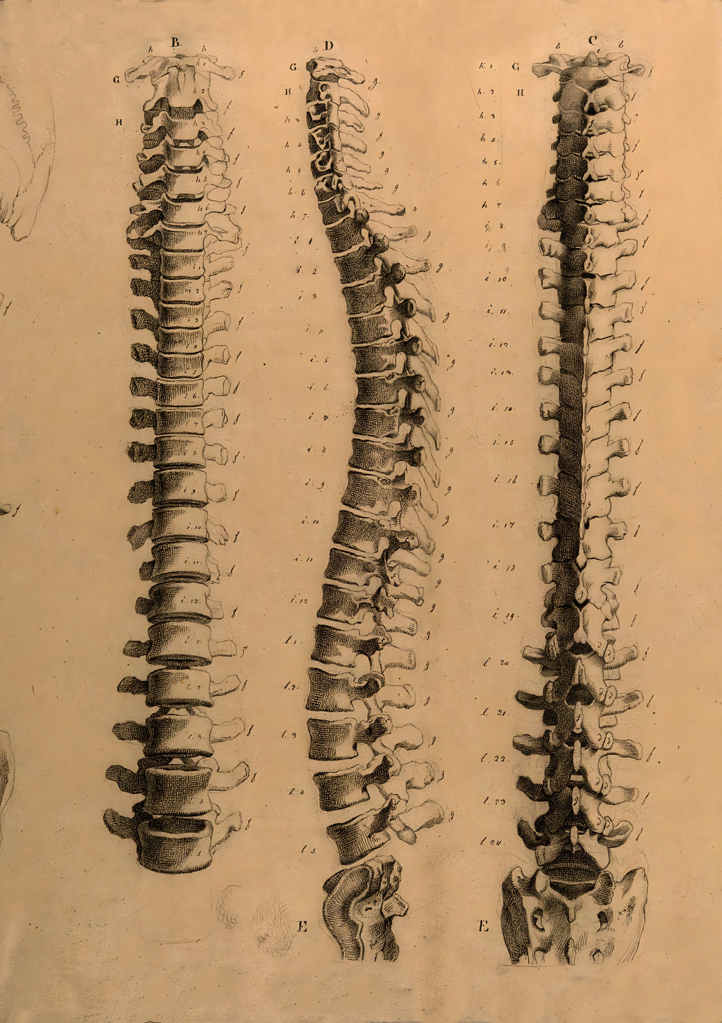
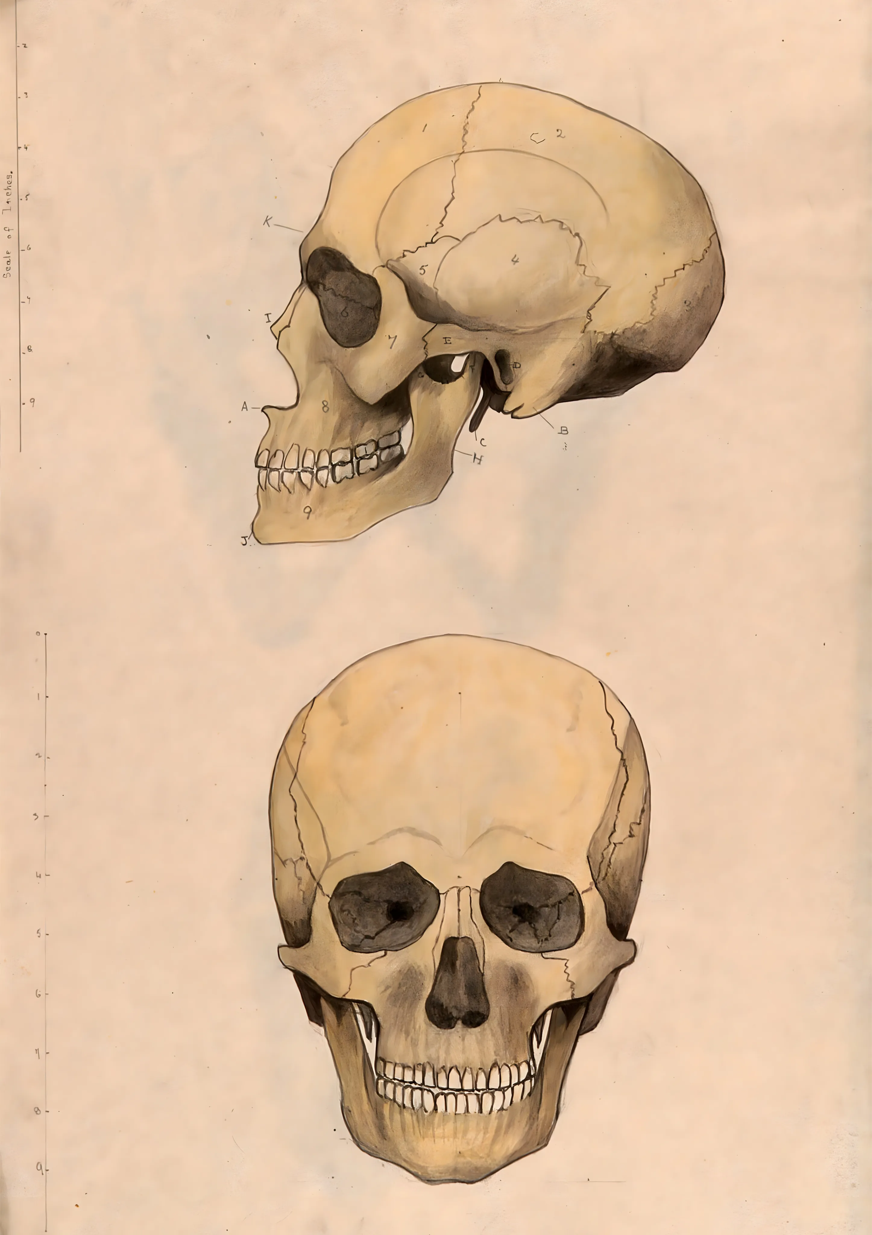
These are anatomical illustrations in the tradition of 19th-century medical plates, reworked as decorative images. They show the heart, brain, skull, skeleton and spine, the muscles of the back and leg, the eye and the arteries of the forearm, alongside phrenology charts, studies of the foetus in the womb and the “Vitruvian Man”.
Anatomie
FromNow €5.00Was €6.00
Anatomie
FromNow €5.00Was €6.00
Anatomie
FromNow €5.00Was €6.00
Anatomie
FromNow €5.00Was €6.00
Anatomie
FromNow €5.00Was €6.00
Anatomie
FromNow €5.00Was €6.00
Anatomie
FromNow €5.00Was €6.00
Anatomie
FromNow €5.00Was €6.00
Anatomie
FromNow €5.00Was €6.00
Anatomie
FromNow €5.00Was €6.00
Anatomie
FromNow €5.00Was €6.00
Anatomie
FromNow €5.00Was €6.00
Anatomie
FromNow €5.00Was €6.00
Anatomie
FromNow €5.00Was €6.00
Anatomie
FromNow €5.00Was €6.00
Anatomie
FromNow €5.00Was €6.00
Anatomie
FromNow €5.00Was €6.00
Anatomie
FromNow €5.00Was €6.00
Anatomie
FromNow €5.00Was €6.00
Anatomie
FromNow €5.00Was €6.00
Anatomie
FromNow €5.00Was €6.00
Anatomie
FromNow €5.00Was €6.00
Anatomie
FromNow €5.00Was €6.00
Anatomie
FromNow €5.00Was €6.00
Try removing one — the counts beside each option say how many prints it would leave.
Clear allMedical plates of the nineteenth century showed the body one structure at a time, with each vessel, bone and muscle named. The prints here keep that clinical way of looking. Phrenology, founded by Franz Joseph Gall around 1800, claimed to read character from the shape of the skull; the theory is discredited, but its labelled head charts survive. The “Vitruvian Man” and the foetus studies are drawings by Leonardo da Vinci.
The plates group by system: the heart from several sides, the blood vessels of the brain, the skull, spine and thorax, the back muscles, the tendons of the hand. Titles such as “Muskeln des Menschen” and “Encéphale de l’homme” point to German and French sources. Made to order in Berlin, each is a giclée print with pigment inks on matte 210 g/m² fine art paper or 400 g/m² cotton canvas, in sizes A4 to A0.